LEAD: A Mayo Clinic research team has validated an artificial intelligence system that detects pancreatic cancer on routine CT scans more than a year before clinical diagnosis, outperforming expert radiologists by a wide margin in a multi‑institutional study published in Gut.
A Disease That Hides in Plain Sight
Pancreatic ductal adenocarcinoma (PDA) — the most common form of pancreatic cancer — remains one of the deadliest malignancies in modern medicine. More than 85 % of patients receive their diagnosis after the disease has already spread beyond the pancreas, at which point curative surgery is no longer possible. Five‑year survival languishes below 15 % in the United States, and projections from the National Cancer Institute indicate pancreatic cancer will become the second‑leading cause of cancer‑related death in the U.S. by 2030.
The fundamental problem is not that treatments are powerless. It is that the pancreas, tucked deep in the abdomen, can harbour a growing tumour for years without producing symptoms — and when it does, the signs are nonspecific: vague abdominal discomfort, unintended weight loss, new‑onset diabetes. By the time a CT scan reveals a visible mass, the window for curative intervention has usually closed. As lead researcher Dr. Ajit Goenka of the Mayo Clinic put it, “the greatest barrier to saving lives from pancreatic cancer has been our inability to see the disease when it is still curable.” This insight also frames the urgency behind recent treatment advances, including a triple‑therapy approach that has shown promise against advanced disease.【Internal link candidate: pancreatic cancer triple therapy】
Current screening policy reflects this diagnostic paralysis. The U.S. Preventive Services Task Force (USPSTF) recommends against screening for pancreatic cancer in asymptomatic adults — a Grade D recommendation — because no existing modality (imaging or biomarker) has demonstrated sufficient accuracy to justify population‑level screening without causing harm from false positives and unnecessary invasive procedures. It is precisely this evidence vacuum that the new Mayo Clinic AI model, called REDMOD, is designed to fill.
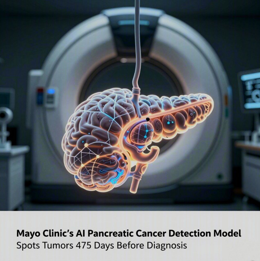
How REDMOD Sees What Radiologists Cannot
REDMOD — short for Radiomics‑based Early Detection Model — is not looking for a tumour. It is looking for the tissue‑level disruption that precedes tumour formation. The model extracts hundreds of quantitative imaging features — texture patterns, intensity gradients, wavelet‑filtered structural signatures — that describe the microscopic architecture of the pancreas on standard abdominal CT scans. These “radiomic” features capture biological changes that occur as cancer begins to develop, long before any focal mass becomes visible to the human eye.
The research team, led by Dr. Sovanlal Mukherjee and senior author Dr. Ajit Goenka at Mayo Clinic in Rochester, Minnesota, trained REDMOD on a multi‑institutional cohort of 969 patients (156 pre‑diagnostic PDA cases and 813 controls) and validated it on an independent test set of 493 patients with a low‑prevalence case mix — 63 pre‑diagnostic cancers versus 430 controls, a roughly 1:6 ratio designed to approximate real‑world clinical settings. The model couples fully automated AI‑driven pancreas segmentation with a heterogeneous ensemble architecture (logistic regression, random forest, XGBoost), eliminating the need for time‑intensive manual outlining while reducing variability.
The headline results, published on 28 April 2026 in Gut, a BMJ specialist journal: REDMOD achieved an area under the curve (AUC) of 0.82, with 73.0 % sensitivity for detecting occult PDA — nearly double the 38.9 % sensitivity achieved by two experienced abdominal radiologists reviewing the same scans (p < 0.001). At lead times greater than 24 months before clinical diagnosis, the advantage widened to nearly threefold: 68.0 % vs. 23.0 %. The median detection lead time was 475 days, or roughly 16 months. Specificity — the model’s ability to correctly identify disease‑free scans — was 81.3 % in a multi‑institutional validation cohort and 87.5 % in the public NIH‑PCT dataset.
Crucially, the model demonstrated longitudinal stability. When the same patients were scanned at different time points, REDMOD’s predictions remained 90–92 % concordant, meaning the signal it detects is not an artefact of scanner variability, patient positioning, or transient inflammation but a genuine, persistent biomarker of early malignant transformation.
Why This Matters: The Arithmetic of Early Detection
The clinical significance of a 475‑day lead time is difficult to overstate. Pancreatic cancer progresses rapidly once it becomes clinically apparent. The median survival for metastatic disease is measured in months, not years. Modelling studies cited by the research team indicate that shifting the proportion of patients diagnosed with localized (resectable) disease from the current 10 % to 50 % would more than double overall survival rates.
REDMOD is designed to integrate into existing clinical workflows, not replace them. The model analyzes CT scans already obtained for other clinical reasons — particularly in high‑risk patients, such as those with new‑onset diabetes and unexplained weight loss, a group for whom the UK’s National Institute for Health and Care Excellence (NICE) already recommends urgent imaging. By flagging elevated risk before a visible mass appears, the AI could serve as a triage tool that prompts closer surveillance or more definitive testing, without requiring additional scans or invasive procedures at the point of screening.
The study does not claim that REDMOD is ready for clinical deployment. The authors are explicit: “prospective validation in high‑risk cohorts” is the necessary next step before the model can be considered for real‑world use. But the evidence base now exists — peer‑reviewed, externally validated, and benchmarked against human performance — to justify those prospective trials.
Frequently Asked Questions
Can AI detect pancreatic cancer early?
Yes, in a research setting. The Mayo Clinic’s REDMOD AI model demonstrated the ability to identify the “invisible” radiomic signature of pancreatic cancer on routine CT scans a median of 475 days — roughly 16 months — before clinical diagnosis. The model achieved 73 % sensitivity, nearly double that of expert radiologists (39 %), and nearly triple for cases detected more than two years before diagnosis. However, the model is not yet approved for clinical use; prospective validation studies are still required.
How does REDMOD differ from a radiologist reading a CT scan?
REDMOD analyzes quantitative imaging features — micro‑scale texture patterns, intensity gradients, and wavelet‑filtered structural signatures — that describe tissue architecture at a level below human visual perception. Radiologists look for visible masses or focal abnormalities. REDMOD detects the diffuse, subvisual tissue disruption that precedes mass formation. The model also automates pancreas segmentation, removing the variability of manual outlining. Of its selected radiomic features, 90 % were wavelet‑filtered, capturing multi‑scale textural disruptions invisible to the naked eye.
Is pancreatic cancer screening recommended for the general population?
No. The U.S. Preventive Services Task Force (USPSTF) currently recommends against routine pancreatic cancer screening in asymptomatic adults — a Grade D recommendation — because no existing test (imaging, blood biomarkers, or otherwise) has proven sufficiently accurate to justify population‑level screening. The balance of potential benefit versus harm from false positives and unnecessary invasive procedures has, until now, been unfavourable. Tools like REDMOD may eventually change that calculus, but only after prospective clinical trials demonstrate real‑world effectiveness.
Editor’s Analysis
Deep Reflections: What This Reveals About the Architecture of Medical Knowledge
Beyond the headline numbers, the REDMOD study exposes something fundamental about how medical knowledge is constructed — and constrained — by the limits of human perception. For decades, the diagnostic paradigm for pancreatic cancer has been governed by what a radiologist can see. A “normal” CT scan was, by definition, a scan in which no mass was visible. This study demonstrates that “normal” was an attribution of perceptual limitation, not biological reality. The cancer was already there; the information was already in the pixels. Human vision simply could not decode it.
This is not a failure of radiology. It is a recognition that pattern recognition at scale — across hundreds of textural features, across time, across institutions — is a computational problem, not a visual one. The REDMOD framework represents a shift from subjective visual interpretation to quantitative tissue phenotyping. And it arrives at a moment when the broader project of AI‑augmented medicine is being forced to move beyond demonstrations of technical capability and into the harder terrain of clinical validation, regulatory clearance, and equitable deployment.
Critical Analysis: How Solid Is the Evidence?
The study warrants cautious optimism, not uncritical celebration. The evidence base is strong for a retrospective validation study, but it has important limitations that the authors themselves acknowledge.
On the positive side: the sample is multi‑institutional, the case–control ratio simulates low‑prevalence real‑world settings, the model is externally validated against two independent cohorts, longitudinal stability is demonstrated, and performance is directly benchmarked against human radiologists. The use of an ensemble architecture and the ablation analysis showing that filtered radiomic features outperform unfiltered features by a statistically significant margin (AUC 0.82 vs. 0.74; p = 0.007) adds mechanistic credibility.
However, the study is retrospective. The pre‑diagnostic scans were identified after cancer was already confirmed histopathologically, which introduces potential selection and ascertainment biases that prospective designs are designed to eliminate. The model was not tested across different ethnic and racial groups, an omission that matters for any tool that might eventually be deployed in diverse populations. The radiologist comparators had only three years of post‑residency experience — not novice, but not the senior subspecialists who might perform differently. And the specificity of 81–87 %, while reasonable, means that in a truly low‑prevalence screening population, the positive predictive value would drop substantially, generating false positives that would need clinical pathways to resolve without causing harm.
Perhaps most importantly, the study demonstrates detection — not mortality reduction. The critical evidence gap between these two endpoints must be closed by prospective trials that randomize high‑risk patients to AI‑augmented screening versus standard care and measure whether earlier detection actually translates into longer survival, fewer emergency presentations, or better quality of life. The history of cancer screening is littered with biomarkers and imaging tools that detected more cancer without saving more lives.
Cui Bono: Who Benefits From This Story?
Several stakeholders stand to gain substantively from the REDMOD narrative, and being explicit about this does not denigrate the science — it contextualizes it.
Mayo Clinic strengthens its positioning as a leader in AI‑augmented radiology at a moment when academic medical centres are competing fiercely for talent, funding, and prestige in the digital health space. The senior author, Dr. Goenka, and the team gain academic visibility and grant‑attracting credentials. BMJ and Gut benefit from publishing a high‑impact paper with broad media appeal.
The radiology AI industry — companies building FDA‑cleared imaging tools — gains a powerful proof‑of‑concept that can be cited in investor decks, regulatory submissions, and marketing collateral, even though REDMOD itself is not a commercial product. The broader narrative that “AI sees what doctors miss” is worth billions in valuation to this sector.
Patients and patient advocacy organizations gain something less transactional but more consequential: hope grounded in evidence. Pancreatic cancer advocacy groups have long argued that the disease’s lethality is as much a failure of detection as of treatment. REDMOD provides them with a concrete, peer‑reviewed milestone to point to when arguing for increased research funding and screening policy reform.
Distraction Analysis: What Bigger Issue Might This Story Crowd Out?
The excitement around an AI model that detects invisible cancer risks obscuring a more uncomfortable truth: even if REDMOD or a successor tool achieves perfect sensitivity and specificity tomorrow, the infrastructure to act on those results does not exist for most of the world’s population — and may not exist for significant portions of the U.S. population either.
Pancreatic cancer surgery is among the most technically demanding procedures in oncology. The Whipple procedure (pancreaticoduodenectomy) carries a perioperative mortality risk of 3–5 % even at high‑volume centres, and outcomes are strongly correlated with surgeon and hospital volume. Detecting more early‑stage cancers in community hospitals that lack high‑volume pancreatic surgery programs would create a referral bottleneck — or worse, lead to surgeries performed at low‑volume centres with poorer outcomes.
Meanwhile, the fact that the USPSTF still recommends against any pancreatic cancer screening reflects a deeper systemic problem: the evidence standards for screening policy rightly require proof of mortality benefit from randomized trials, yet no funder — public or private — has been willing to mount the large, lengthy, expensive trials required to generate that proof for a disease with relatively low incidence. AI models like REDMOD may accelerate the technical capability for early detection without accelerating the political will to fund the definitive trials that would change screening guidelines.
Who Does This Not Serve?
A tool built and validated at Mayo Clinic, on predominantly U.S. patient populations, using CT scanners and imaging protocols common in high‑resource settings, does not automatically serve patients in low‑ and middle‑income countries where CT access is limited, radiologist expertise is scarce, and pancreatic cancer may not even rank among the top health priorities competing for scarce resources. The global health equity implications of AI‑augmented cancer screening are rarely discussed in the press releases, but they are material.
Even within wealthy countries, the populations at highest risk for pancreatic cancer — including Black Americans, who have a roughly 20 % higher incidence than white Americans, and individuals with long‑standing type 2 diabetes — are often the same populations that face barriers to accessing the very CT scans on which REDMOD depends. An AI model that requires a CT scan to function cannot close the gap between those who get scanned and those who do not.
Finally, the radiologists who participated as comparators in this study — junior attendings with three years of experience — may reasonably feel that the comparison undersells human performance at its best, even as the broader narrative frames the result as “AI beats doctors.” The more productive framing — one the study authors themselves lean toward — is not replacement but augmentation: AI as a safety net that catches what even experienced eyes miss. But the media coverage does not always make that distinction, and the professional identity of radiologists — already navigating the most AI‑disrupted specialty in medicine — deserves more careful handling than sensational headlines tend to provide.
Key Takeaways
- Mayo Clinic’s REDMOD AI detected visually occult pancreatic cancer a median 475 days before clinical diagnosis, with 73 % sensitivity versus 39 % for radiologists — a near‑doubling of detection performance at the pre‑clinical stage.
- The model is fully automated, longitudinally stable, and externally validated across multiple institutions, but remains a research tool that requires prospective clinical trials before any real‑world deployment.
- Pancreatic cancer’s lethality is primarily a detection failure: >85 % of cases are diagnosed at an incurable stage, and no population screening programme currently exists. REDMOD targets that gap directly.
- The gap between technical capability and clinical infrastructure — surgical capacity, screening policy, health equity — remains wide and is not narrowed by AI alone.
Internal Links Used
- Advances in pancreatic cancer treatment — placed in “A Disease That Hides in Plain Sight” — contextualizes why early detection matters alongside treatment innovation for the same disease.
- AI cancer diagnosis with electronic nose technology — placed in “How REDMOD Sees What Radiologists Cannot” — illustrates a parallel AI‑driven cancer detection approach, strengthening the broader narrative.
- The morality of scientists and science for sale — placed in “Cui Bono” — deepens the editorial analysis of institutional incentives in biomedical research.
Sources
- Next-generation AI for visually occult pancreatic cancer detection — Gut (BMJ) — peer‑reviewed primary study; open access; published 28 April 2026 — primary / peer‑reviewed
- Mayo Clinic AI helps specialists detect pancreatic cancer up to 3 years before diagnosis — Mayo Clinic News Network — official institutional press release — official
- AI model detects very early normally ‘invisible’ tissue changes of pancreatic cancer — BMJ Group — publisher press release with expert summary — official
- Can a Radiomic AI Model Facilitate Earlier CT Detection of Pancreatic Ductal Adenocarcinoma? — Diagnostic Imaging — independent specialist analysis; 30 April 2026 — high‑credibility reporting
- AI algorithm beats radiologists at spotting early pancreatic cancer — AuntMinnieEurope — radiology trade publication; 28 April 2026 — high‑credibility reporting
- USPSTF Pancreatic Cancer Screening — Screening for Pancreatic Cancer (ScienceDirect summary) — evidence‑based screening guideline context — authoritative reference